Induced Electric Fields in Patients (MRI)
Description:
Controlled E-field gradient coils
Summary
A novel magnetic coil for MRI Scanners was developed by Sir Peter Mansfield and Prof. Roger Bowley at the University of Nottingham. The new gradient coil was designed to enable higher resolution in MRI scanners but at a reduced peripheral nerve stimulation in patients.
Key Benefits
· The technology will enable the new high-field MRIs to conform to European regulations which limit workers’ acute exposure to strong electromagnetic fields.
· The technology would enable high-field MRI systems to generate higher fields safely without compromising on performance.
· The technology would both increase MRI resolution and reduce the side effects that the higher resolution would bring.
· The new technology fits in with current designs and can be retrofitted to standard MRI machine design.
  Gradient coil– 30 m of 6 x 3 mm 2 X-section per coil.
Markets Sectors
This technology addresses and caters for companies developing high-field magnetic system for high resolution imaging. High-field, high resolution systems are reported to represent the fastest growing segment; hospitals and clinics upgrade old equipment with state-of-the-art systems.
Patient comfort is a major factor in this market. The effect of high magnetic field on both workers and users is also a major concern. This is particularly important with the higher resolution machines. A European directive was passed to address this issue making it difficult to use the traditional style magnetic coil for high field applications in Europe. This technology will help gradient coil designers to use higher field for high resolution image, but also patient comfort.
Technical Information
Peripheral neural stimulation is a major problem in traditional gradient coil designs. Induced current problems in patients relate directly to gradient strength and modulation frequency. Current designs of gradient coil tend to limit ultra-high-speed imaging methods such as EPI (echo-planar imaging) as the induced currents would produce tingling sensations and involuntary muscle twitch. Neural stimulation could also trigger epileptic fits and/or cardiac fibrillation.
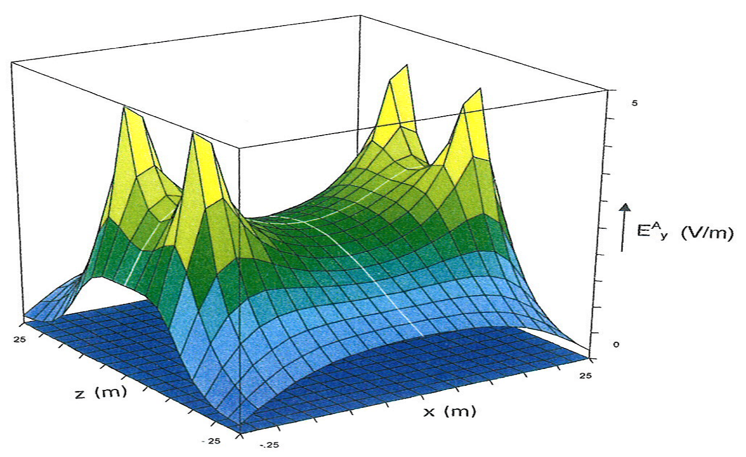
An important aspect for reduction of induced currents is the coil geometry. It is desirable to design the gradient coil in such a way as to prevent closed loop circulating currents within the body. Preliminary results using a four-sector gradient coil with rectangular geometry, operating in a low mutual coupling mode, indicate significant reduction in the E-field within the subject volume of the coil. Reduction in induced currents in the patient allows safer operation at higher magnetic field strengths together with faster scans currently prohibited through neural stimulation effects in standard coil geometries.
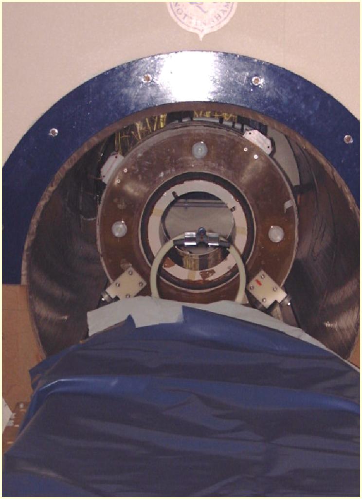
The magnetic gradient coil, which incorporates active acoustic control to give 40dB noise reduction over the object field, was used to produce a snap-shot image of a phantom in 10.6mS using EPI operation at 3.0 T as shown below.
IP StatusThis technology is currently protected by an international patent - published as WO 2003/038461.
Gradient coil– 30 m of 6 x 3 mm 2 X-section per coil.
Markets Sectors
This technology addresses and caters for companies developing high-field magnetic system for high resolution imaging. High-field, high resolution systems are reported to represent the fastest growing segment; hospitals and clinics upgrade old equipment with state-of-the-art systems.
Patient comfort is a major factor in this market. The effect of high magnetic field on both workers and users is also a major concern. This is particularly important with the higher resolution machines. A European directive was passed to address this issue making it difficult to use the traditional style magnetic coil for high field applications in Europe. This technology will help gradient coil designers to use higher field for high resolution image, but also patient comfort.
Technical Information
Peripheral neural stimulation is a major problem in traditional gradient coil designs. Induced current problems in patients relate directly to gradient strength and modulation frequency. Current designs of gradient coil tend to limit ultra-high-speed imaging methods such as EPI (echo-planar imaging) as the induced currents would produce tingling sensations and involuntary muscle twitch. Neural stimulation could also trigger epileptic fits and/or cardiac fibrillation.
An important aspect for reduction of induced currents is the coil geometry. It is desirable to design the gradient coil in such a way as to prevent closed loop circulating currents within the body. Preliminary results using a four-sector gradient coil with rectangular geometry, operating in a low mutual coupling mode, indicate significant reduction in the E-field within the subject volume of the coil. Reduction in induced currents in the patient allows safer operation at higher magnetic field strengths together with faster scans currently prohibited through neural stimulation effects in standard coil geometries.
The magnetic gradient coil, which incorporates active acoustic control to give 40dB noise reduction over the object field, was used to produce a snap-shot image of a phantom in 10.6mS using EPI operation at 3.0 T as shown below.
IP StatusThis technology is currently protected by an international patent - published as WO 2003/038461.
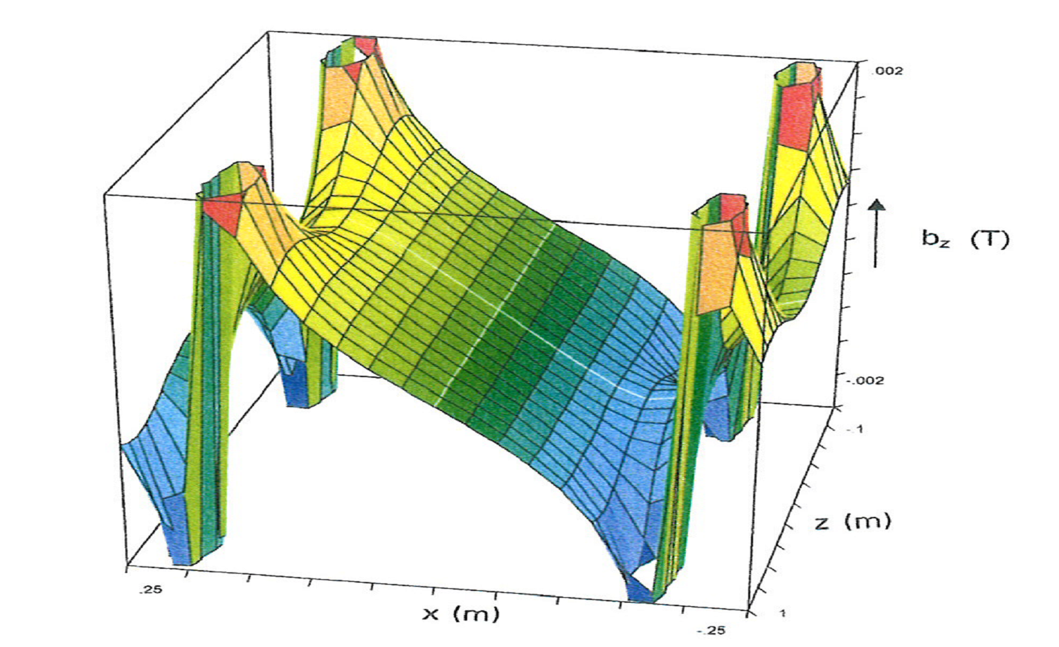 Magnetic field bz produced by the gradient coil when the plates for the modified four-sector gradient coil are shorted and electric field between the plates is uniform across the plate diameter.
 Three-dimensional plot of the spatial distribution of the induction field for the coil arrangement.
Title
|
(EN) MRI GRADIENT COILS WITH REDUCED NEURAL STIMULATION
|
Abstract:
|
(EN) A novel approach to the design of gradient coils for MRI is introduced which takes into account from the start the effects of induced E-fields and hence currents in a patient subjected to time dependent magnetic field gradients. The approach has led to conceptually novel designs of gradient coils which comprise distributions of electrodes or tessellae placed around the basic coil structure. When properly energised the electrode arrays are able, in the simplest case, to reduce the maximum E-field experienced by the patient by approximately a factor of three over that experienced with a standard fingerprint or distributed transverse gradient coil, the comparison being done for the same gradient strength, same coil diameter and same ROI.
|
| | |
Inventors:
Keywords:
|


No comments:
Post a Comment